
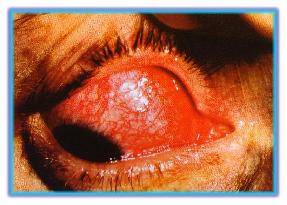
| Sckeritis can be
classified as anterior or posterior. Anterior scleritis
is divided into sectoral, diffuse, nodular and
necrotizing subtypes. Patients present with deep,constant
pain in the involved eye. The disease can be bilateral in
50% of patients. It occurs predominantly in women and
varies from mild forms to very severe forms that can
destroy the eye. With active inflammation,the sclelra has
a violaceous hue best seen lin naturnal sunlight.
Engorgement of the deep vascular plexus is evident,as
shown in the slide;the engorgned vessels do not move when
the overlying tissues are moved with a cotton-tipped
applicator. Nodular anterior scleritis is most commonly
seen in association with rheumatoid arthritis. The more
destructive necrotizing scleritis can be associated with
substantial loss of vision. The patient's life expectancy
has been shown to be adversely affected by uncontrolled
associated systemic autoimmune disease. Posterior scleritis almost always presents with pain,tenderness,and proptosis. Choroidal folds,exudaive retinal detachment and papilledema van also occur. Complications of scleritis include Keratitis, uveitis, cataract, glaucoma, scleral thining, marginal Keratolysis and scleral perforation. The disease can occur as an isolated phenomenon or in association with various systemic autoimmune diseases, such as rheumatoid arthritis, Wegener's granulomatosis, polyartheritis nodosa, systemic lupus erythematosus, herpes zoster ophthalmicus, syphilis, tuberculosis and gout. Milder cases respond to topical steroid therapy. Although definitive treatment varies according to cause,treatment sometimes involves nonsteroidal anti-inflammatory agents, systemic steroids, and, for the progressive cases, immunosuppressive aggents. Appropriate treatment of associated systemic autoimmune diseases is also important. |
 |
Previous |
Scleritis | Episcleritis | Scleromalacia Perforans | Congenital Melanosis Oculi
Eye Lid | Lacrimal System Disorders | Scleral Disease